A woman tucks a violin under her chin, takes a deep breath, and draws a bow across the strings. The camera circles her, taking in her performance of Bach’s Sonata No. 1 in G Major from all angles — her fingers dancing on the instrument’s neck, her upper body swaying with the music.
Then, just before the viewer succumbs completely to the music’s spell, another figure materializes in the background. He has an academic, nerdy-chic look about him — bow tie, black jacket adorned with red handkerchief, and thick, round spectacles.
With professorial authority, he intones: “We humans have a unique ability to interact with the world around us using our upper limbs. Precise motor maps created in the brain enable us to move specific muscle groups in discrete ways.”
“These are not your typical educational videos,” explains Professor Wayne Vogl, the nerdy-chic co-star in a set of 14 short neuroanatomy videos created by the UBC Faculty of Medicine, the brainchild of Claudia Krebs, a Professor of Teaching in the Department of Cellular and Physiological Sciences.
Telling a story for greater understanding
“I’ve watched videos of anatomists teaching anatomy, and it’s usually very dry: ‘This is the artery, this is the nerve, this is the muscle,’ ” Dr. Vogl says. “There’s no sense that there’s a bigger story to tell. We wanted to show that the upper limb’s anatomy is designed to position your hand in space to use as a sensory and motor tool. A student starting from that point would be in a better position to build into it all of the detail they need.”
Dr. Krebs produced the videos for UBC students, but they are now being viewed by students around the world – sometimes because their instructors have required it.
“This has been a career-defining project for me, and it has led to connections with colleagues around the world,” Dr. Krebs says.

Claudia Krebs with students in the “Brain and Behaviour” lab. Photo by Martin Dee
The videos began as part of a larger project undertaken by Dr. Krebs to transform the “Brain and Behaviour” block she taught for more than a decade.
Inspired by the trend toward “flexible learning,” she wanted to provide her students with more content before lectures and labs, so that classroom time could be re-oriented toward analysis and application to clinical cases. But to entice students to dive into such complex material on their own, she knew it had to be interactive, engaging, even stylized.
But Dr. Krebs didn’t find anything that reflected UBC’s distinctive approach to teaching anatomy, which emphasizes how everything fits together into a larger whole. Nor did she find anything that was very watchable.
So she teamed with Dr. Vogl, Zac Rothman, a video producer in the Faculty’s educational technology office, and a few eager graduate students to make some material of their own.
A debut, and then a sequel
The first set of nine videos, funded with a grant by UBC’s Flexible Learning initiative, was released in 2014. Anecdotally, Dr. Krebs noticed greater confidence among her students. That observation was confirmed by exam results from the first two cohorts: the bell curve distribution of grades shifted to the right, with the lowest-scoring students moving from the 60s to the 70s.
“Teaching is not so much about knowledge transmission, as it is about helping students understand,” Dr. Krebs says. “These videos set the tone by focusing on how things work, and how they fit into the larger whole, and made for more vivid class discussions.”
So, like any self-respecting producer, she decided to make a sequel – or five. They picked up where the first series left off, focusing on extensions of the central nervous system to rest of the head, as well as the arm.
“In the second series, we had a lot more fun with it, because we knew the medium, and we played with it more,” Dr. Krebs says.
Each video started with scripts that were initially written by Dr. Krebs or Dr. Vogl, then refined and pared down by the team in “writers’ room” sessions, where every line was scrutinized and revised for clarity, coherence, even eloquence.
“We wanted the words to roll off the speaker’s tongue,” Dr. Krebs says.
A sublime cinematic moment
Beyond the words, the team would brainstorm for the visual hooks that would embed the concept in the viewer’s mind, and elevate the videos to something akin to art.
“We tried to have those memorable moments so people could connect with it,” she says.
For the brachial plexus video, the team’s original idea was to film young children playing with toys. After concluding that using children would be too challenging, they hit upon the idea of playing the violin, which depends on movement of the upper limb.
That is how Jennie Press, a violinist in the Vancouver Symphony Orchestra, found herself performing Bach in a most unlikely venue: UBC’s immense and immaculate gross anatomy lab, where medical students dissect donated cadavers. (For the shoot, the covered bodies were pushed off to the side, outside the frame.)
For a smooth tracking shot, Rothman mounted a camera on a circular rail surrounding the musician. And Dr. Vogl, after several attempts, mastered the multi-tasking – the walking, talking and looking – required for that opening sequence.
“The whole thing was just spectacular,” Dr. Krebs says. “It almost brought tears to my eyes.”
Make ‘em laugh – and they’ll remember

A Monty Pythonesque scene from one of the neuroanatomy videos.
In addition to the sublime, they also indulged in the ridiculous.
For an overview of the head and neck, Rothman – inspired by diagrams Dr. Vogl created for Gray’s Anatomy for Students – proposed using Monty Pythonesque animation. So Dr. Vogl was transformed into a bespectacled, bow-tie clad avatar.
Playfulness – more than one would ever expect in an anatomy lesson – abounds. To convey how systems for breathing and systems for eating or drinking are connected and separated by a system of valves, graduate student Arlo Adams downs a glass of water, holds it in his mouth, and dramatically inhales and exhales through his nose to the beat of a raucous drum solo.
The video concludes with interviews of children from Lord Tennyson Elementary School recounting instances of drinking and laughing at the same time, with the inevitable result: a spray of liquid shooting out of the nose.
“It just kind of went everywhere,” one girl says.
“It brought comic relief to a content-heavy video,” Dr. Krebs says. “And if you laugh at something, you remember it better.”
The start of something bigger
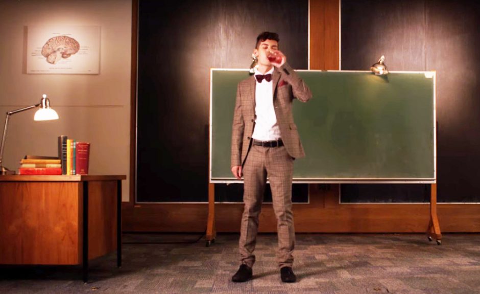
Graduate student Arlo Adams dramatically downs a glass of water to demonstrate how systems for breathing and systems for eating or drinking are connected and separated by a system of valves.
From the start, Dr. Krebs was determined to share these videos far beyond UBC. So she posted them on YouTube, and without any marketing or buzz, they slowly went viral – at least by the standards of medical education videos.
The first installment, “Introduction to the Central Nervous System,” has been viewed close to 150,000 times. The medical schools of Boston University, the University of Washington, the University of the West Indies, the University of Ottawa and Vrije Universiteit (“Free University”) Amsterdam have added the videos to their curricula. Students from Hawaii, Louisiana, the U.K., Australia, Germany, Bangladesh, Brazil and even Iraq have emailed their praise. An online search for “neuroanatomy” brings up the landing page for Dr. Krebs’ course material – at the very top of the results.
“I saw the first two, and I immediately sent an email to her to congratulate her, because they were the best-shot educational videos I have ever seen,” said Jochen Bretschneider, a lecturer at Vrije Universiteit’s School of Medical Sciences and an ear, nose and throat surgeon at its affiliated medical centre. “Very nice light, very nice focus, very nice audio. They were short, to the point.”
Dr. Bretschneider, who recently earned a filmmaking degree, has produced some of his own medical education videos. With Dr. Krebs’ permission, he included UBC’s work in Vrije Universiteit’s collection.
The videos have brought together like-minded anatomists around the world, in Amsterdam, Sweden, Glasgow and Auckland, who are creating similar material – not just videos, but three-dimensional interactive models. Their working name: the Anatomy Hub.
“There’s no point in the guys in the Netherlands doing their own video on the brachial plexus. It’s done, they can go ahead and use it,” Dr. Krebs says.
Taking a stand for anatomy
She and her colleagues aim to create a repository of anatomy material that would help preserve anatomy’s footprint in an ever-evolving medical curriculum.
“The basic science instructors are realizing that we need to get our information out there to students, because some of it is being pushed to the side,” Dr. Krebs says. “This is a good way to ensure the students get that content, even if it’s only piecemeal, with students seeking it out when they feel they need it.”
She and her colleagues also believe this kind of material can be particularly helpful for medical students in developing countries, where qualified instructors are in short supply. A three-time winner of teaching awards from medical students and the Faculty of Medicine’s first Professor of Teaching, Dr. Krebs would never claim that videos can replace in-person instruction. But digital resources like these, if educationally sound and expertly produced, can make the educational experience so much better, because they free up classroom time to put content into context.
“UBC, as one of the best universities in the world, has a social responsibility to reach out,” she says. “We can’t just stay in our ivory tower and keep our knowledge and expertise to ourselves. Not every university has the resources to create educational material like this. We do. For us to share that knowledge is part of our responsibility and part of where education, globally, is going. And UBC can be leader with that.”